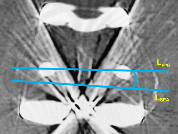
Figure 4.

Method for measuring femoral component rotation using computed tomography. Femoral component rotation was evaluated on the axial slice where two femoral pegs and the medial and lateral epicondyles were visible. Femoral rotation was the angle between the line connecting the two pegs (L peg) and the surgical epicondylar axis (L SEA). The rotational position was classified into three groups: internal rotation from the surgical epicondylar axis (I), 0–3° external rotation (N), and >3° external rotation (E). SEA, surgical epicondylar axis.
