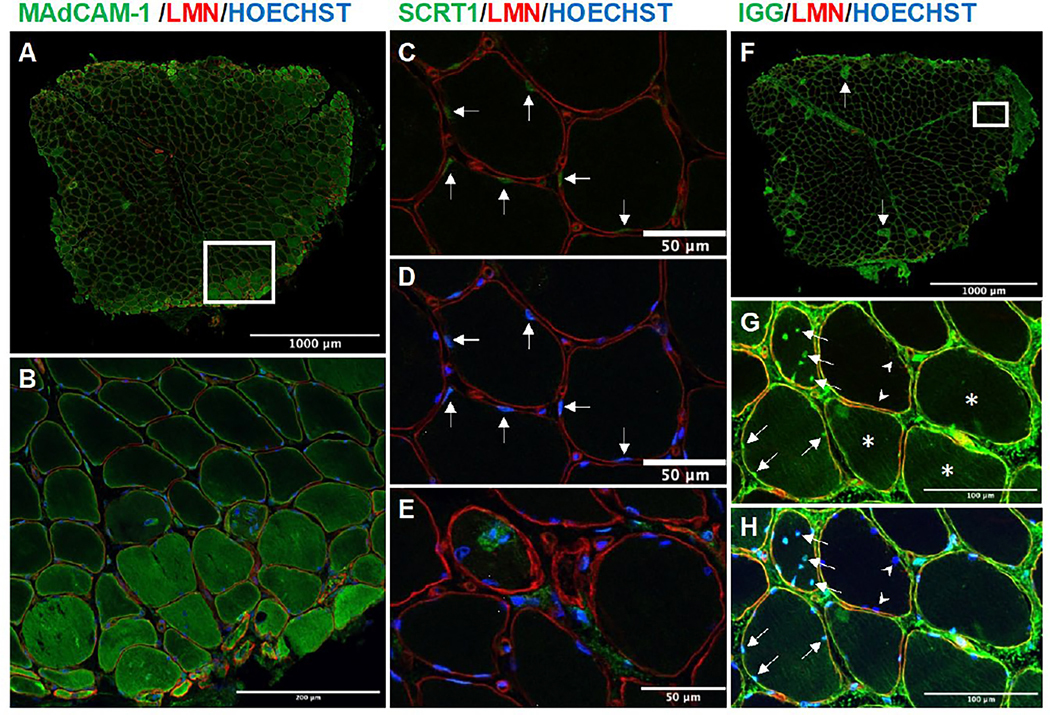
Figure 7.

Confocal microscopy images of the muscle biopsy corresponding to panel 6B showing immunofluorescence staining of MAdCAM-1 (A-B), SCRT1 (C-E), and human immunoglobulin (F-H) co-stained with laminin and Hoechst. MAdCAM-1 was predominantly expressed in the perifascicular region (A and B), with varying levels in different muscle fibers (B). SCRT1 was primarily expressed in the nuclei of muscle fibers (arrows C [substracted Hoechst] and D), in the cytoplasm surrounding the nuclei of severely damaged fibers (E). Human immunoglobulin deposition was most prominent in muscle fibers that were MAC-positive (arrow in F, corresponding MAC staining in Supplementary Figure 5). However, healthy-looking muscle fibers showed lower levels of immunoglobulin deposition (asterisks in G). Notably, a significant number of cell nuclei displayed evidence of immunoglobulin deposition, even in cases where the cytoplasmic deposition was absent (indicated by the arrow in G [subtracted Hoechst] and H). Of note, adjacent fibers did not exhibit any staining for nuclear immunoglobulin, as indicated by the arrowheads in G and H. Supplementary stainings for this biopsy are included in Supplementary Figure 5 and the individual channels of each region of interest are included in Supplementary Figure 6. B was obtained from the region marked with a square in A, and G-H was obtained from the region marked with a square in F.
