. 2024 Dec 2;13:RP98449. doi: 10.7554/eLife.98449

Figure 3. Glycolysis is required for IFN-γ induced expression of activation markers by MDM and not AM.

Human AM (A, C, E) isolated from bronchoalveolar lavage fluid. PBMC were isolated from buffy coats and MDM (B, D, F) were differentiated and adherence purified for 7 days in 10% human serum. Cells were left unprimed (black) or primed with IFN-γ (red) or IL-4 (blue) (both 10 ng/ml) for 24 hr. Cells were left untreated (solid) or treated with 2DG (5 mM; empty) 1 hr prior to stimulation with iH37Rv (MOI 1–10; square) or LPS (100 ng/ml; triangle) or left unstimulated (circle). After 24 hr, cells were detached from the plates by cooling and gentle scraping and stained for HLAR-DR (A, B), CD40 (C, D), CD86 (E, F) and analysed by flow cytometry. Each linked data point represents the average of technical duplicates for one individual biological donor (n=8–9). Statistically significant differences were determined using two-way ANOVA with a Tukey post-test (A–F); *p≤0.05, **p≤0.01, p***≤0.001, ****p≤0.001.

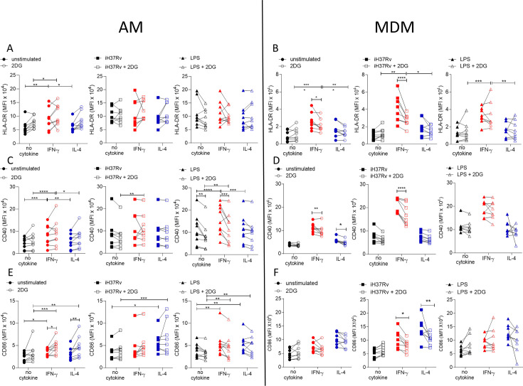
Figure 3.

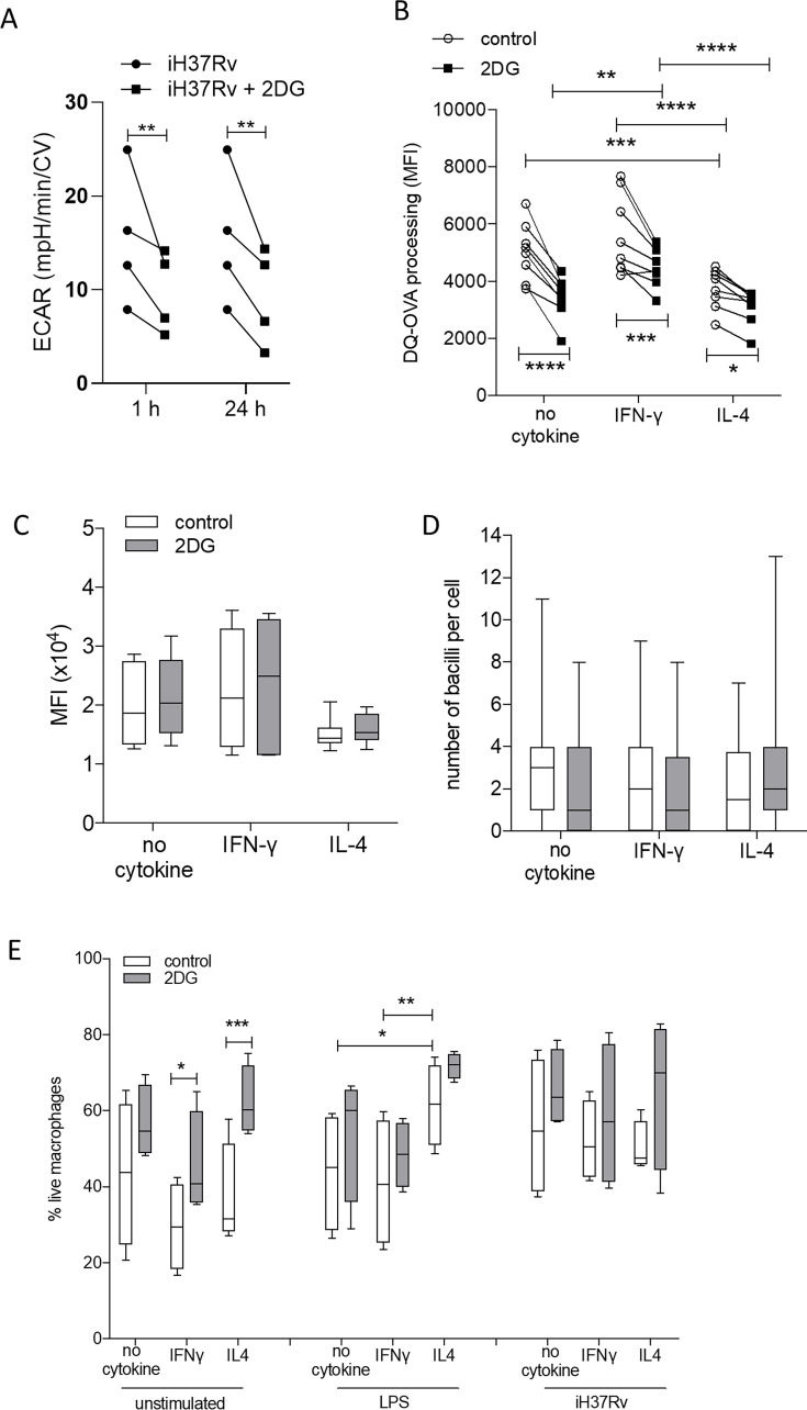
Figure 3—figure supplement 1. 2DG reduces ECAR and antigen-processing but does not reduce phagocytosis or cell viability.

Figure 3—figure supplement 1.

PBMC were isolated from buffy coats and MDM were differentiated and adherence purified for 7 days in 10% human serum. MDM were left untreated or treated with 2DG (5 mM) for 1 or 24 hr. MDM were stimulated with iH37Rv and ECAR was assessed at indicated time points (A). MDM were primed with IFN-γ or IL-4 (both 10 ng/ml) for 24 hr. MDM were treated with 2DG (5 mM) for 1 hr prior to treatment with DQ-Ovalbumin (B; 500 ng/ml; 30 min), fluorescent latex beads (C; 15 min) or iH37Rv (E; MOI 1–10; 180 min). MDM were then detached with ice cold PBS, scraped and analysed by flow cytometry. Alternatively, 1 hr after 2DG MDM were stimulated with iH37Rv (MOI 1–10) or LPS (100 ng/ml) for 24 hr. MDM were then scraped and stained with Zombie NIR for cell viability (E), measured by flow cytometry. Each data point represents one individual donor. Statistically significant differences were determined using two-way ANOVA with a Tukey post-test; *p≤0.05, **p≤0.01, ***p≤0.001, ****p≤0.0001.