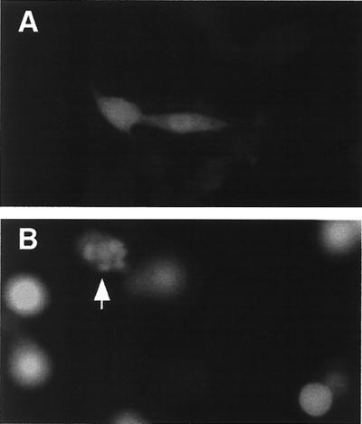
FIG. 1.
M protein expression induces morphological changes associated with apoptosis. BHK cells were transfected with EGPF mRNA (A) or M mRNA and EGPF mRNA (B). At 24 h posttransfection, cells were analyzed by fluorescence microscopy using a 25× water immersion objective. The fluorescein channel was used to detect the presence of EGFP to indicate transfected cells. Digital fluorescence images of transfected cells expressing EGFP are shown in both panels. the arrow in panel B indicates a cell undergoing membrane blebbing.

