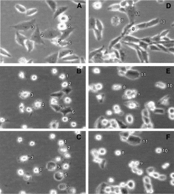
FIG. 4.
Time lapse microscopy analysis of morphological changes in VSV-infected cells. HeLa cells (A to C) or BHK cells (D to F) were infected with wtO virus and then analyzed by phase-contrast time lapse microscopy. Digital images of the same fields captured at different times postinfection are shown. The images of infection of HeLa cells were taken at 4 h (A), 9.5 h (B), and 15.5 h (C). The images of infection of BHK cells were taken at 0.5 h (A), 11 h (B), and 18 h (C). Numbered cells were chosen to illustrate cell rounding due to mitosis (cells 1 and 2), membrane blebbing (cells 3, 4, 8, 9, and 10), and cell blistering due to apoptosis (cells 5, 6, and 7), and a BHK cell that remained elongated at a late time postinfection (cell 11).

