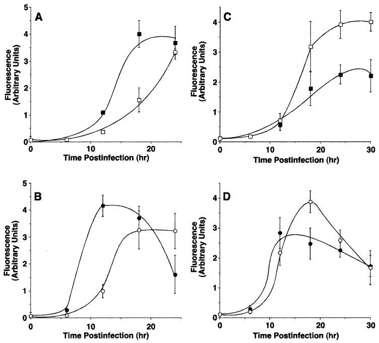
FIG. 6.
Caspase-3 activity in cells infected with wt VSV and VSV M gene mutants. HeLa cells (A and B) or BHK cells (C and D) were infected with rwt virus (closed squares), rM51R-M virus (open squares), wtO virus (closed circles), or tsO82 virus (open circles) for the times indicated. Duplicate samples were analyzed for caspase-3 activation as described in the legends to Fig. 2 and 3. The amount of caspase-3 activated is expressed as arbitrary fluorescence units. The data represent the average ± the standard deviation of three experiments.

