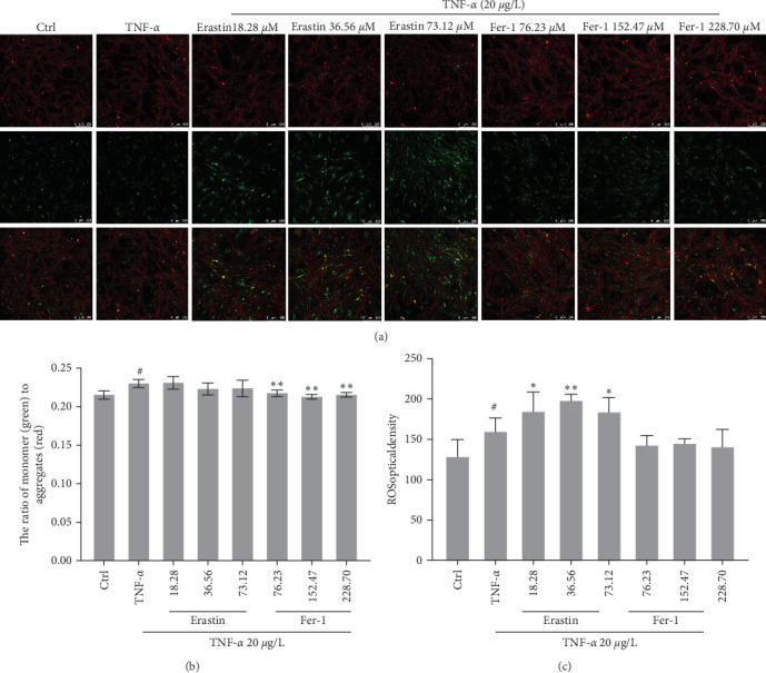
Figure 4.

Ferroptosis impact on mitochondrial membrane potential and reactive oxygen species (ROS) in synoviocytes. About 20 μg/L TNF-α and concentrations gradient of erastin or fer-1 were respectively added in cells to coculture for 48 hr: (a and b) JC-1 company with high mitochondrial membrane potential to accumulate a polymer and produce red fluorescence in the mitochondria matrix. JC-1 cannot aggregate when the mitochondrial membrane potential is lower in the mitochondria matrix. Meanwhile, JC-1 can produce green fluorescence, which facilitates the detection of changes in mitochondrial membrane potential by changes in fluorescence color. It is usual to use the ratio of red–green fluorescence to measure the ratio of mitochondrial depolarization; (c) intracellular ROS was monitored by fluorescence spectrophotometer (#P < 0.05 vs. Ctrl group; ∗P < 0.05 and ∗∗P < 0.01 vs. TNF-α group, n = 5).
