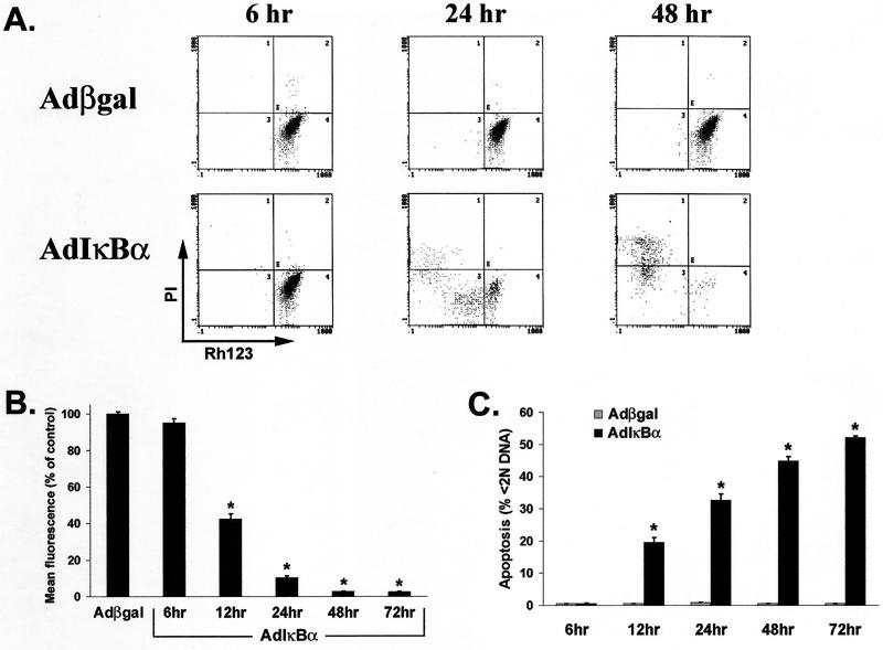
FIG. 5.
Macrophage infection with AdIκBα, but not Adβgal, induces ΔΨm collapse, cell death, and DNA fragmentation in a time-dependent manner. Primary human macrophages differentiated for 7 days were infected with either AdIκBα or Adβgal, and each sample was analyzed for mitochondrial dysfunction, viability, and subdiploid DNA content. (A) AdIκBα-infected macrophages display a time-dependent loss of ΔΨm, assessed by decreased Rh123 fluorescence (x axis), and a subsequent increase in cell death, as determined by incorporation of PI (3 μg/ml; y axis), compared to Adβgal-infected control cells. The data represent one of three replicate samples from a representative experiment. (B) AdIκBα-infected macrophages exhibit reduced Rh123 fluorescence over time compared to those infected with Adβgal. At the indicated times, the cells were incubated with Rh123 (0.1 μg/ml) for 30 min, harvested, and analyzed for Rh123 fluorescence by flow cytometry. The mean fluorescence of Adβgal cultures at each time point represents 100% fluorescence. (C) Infection with AdIκBα, but not Adβgal, increases DNA fragmentation in a time-dependent fashion. Following Rh123 analysis, cells were fixed in 70% ethanol and assessed for subdiploid (<2N) DNA content. The results are representative of five independent experiments. The values in panels B and C represent the mean ± standard error of triplicate cultures. ∗, P < 0.001 of the AdIκBα-infected cells compared to Adβgal infection.

