
Figure 1.
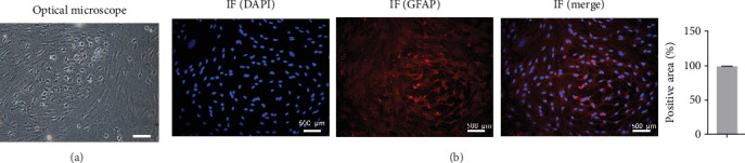
Identification of primary astrocytes using microscopy and immunofluorescence. (a) Cell morphology was observed under an inverted phase-contrast microscope. (b) Detection of glial fibrillary acidic protein expression in an immunofluorescence assay. Scale bar = 500 μm; n = 3.