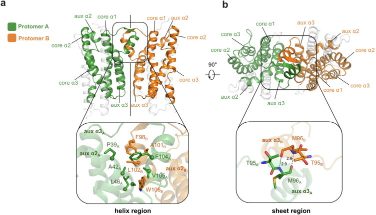
Extended Data Fig. 1. Dimeric structure of AetD and the dimer interface.
a, (top) The dimeric structure of AetD. Part of the auxiliary helix α3 forms the dimerization domain (enclosed by the black the box) that consists of a helix region and a sheet region. (bottom) zoomed-in view of the helix region. Only half of this 2-fold symmetrical dimer interface is shown for simplicity. The helix region is largely hydrophobic, containing hydrophobic residues from the dimerization domain and auxiliary helix α2 from both protomers. b, the dimeric structure rotated 90° out of plane highlighting the sheet region of the dimerization domain. (bottom) zoomed-in view of the sheet region, which is made of two residues from each protomer (T95 and M96) held together by backbone hydrogen bonds.

