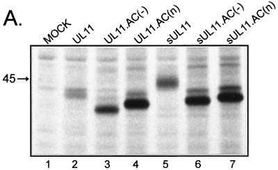
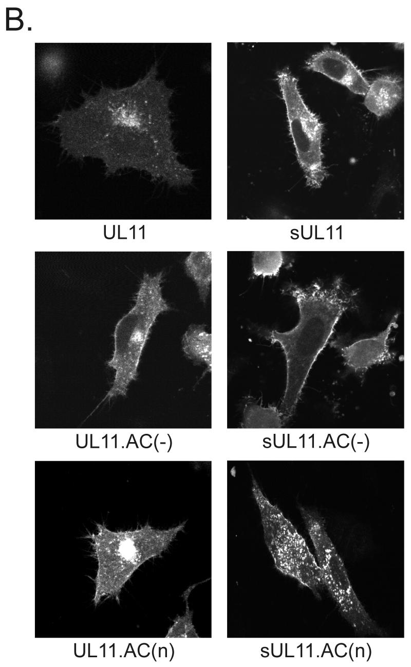
FIG. 2.
Expression of UL11 mutants with inactivated acidic clusters. (A) Biochemical analysis. A7 melanoma cells transfected with the indicated constructs were labeled for 2.5 h with l-[35S]methionine, and UL11-GFP chimeras were immunoprecipitated from cell lysates with a polyclonal antibody specific for GFP. Proteins were separated by SDS-PAGE and visualized by autoradiography. The position of the 45-kDa molecular mass marker is indicated. (B) Subcellular localization. Plasmids were transfected into A7 cells as in panel A, and UL11-GFP chimeras were visualized by live-cell confocal microscopy at approximately 18 h following transfection.


