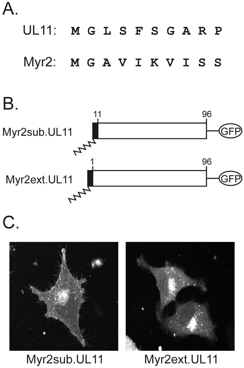
FIG. 6.
Analysis of the first 10 residues of UL11. (A) Sequence comparison. The first 10 amino acids of the UL11 protein and a myristylated form of the Rous sarcoma virus Gag protein are shown. The glycine residue at the second position is the site of myristylation for both proteins, but otherwise the sequences are highly divergent. (B) Myr2-UL11 chimeras. The first 10 residues of the Myr2.Gag protein (black rectangle) and its associated myristate (wavy line) were either substituted in place of the first 10 residues of UL11 (Myr2sub.UL11) or attached to the N terminus as an extension (Myr2ext.UL11). Both constructs have the GFP protein (open oval) fused to their C termini. (C) Subcellular localization. The constructs were transfected into A7 cells, and 18 h later, the UL11-GFP chimeras were visualized by live-cell confocal microscopy.

