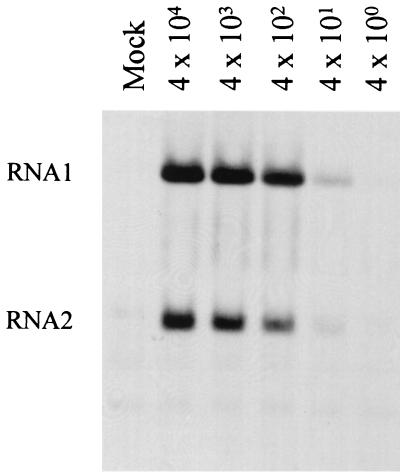
FIG. 1.
Infectivity of PaV in FB33 cells. Helicoverpa zea FB33 cells were infected with 10-fold dilutions of purified PaV (MOIs of 4 × 10⁴ to 4 × 100) or mock infected and incubated at 28°C. After 22 h of incubation, actinomycin D was added at 20 μg/ml, and 30 min later replicating RNAs were metabolically labeled by incorporation of [3H]uridine (20 μCi/ml) for a period of 2 h before total cellular RNAs were harvested. RNAs were resolved by electrophoresis on a 1% agarose-formaldehyde gel and visualized by fluorography. PaV genomic RNA1 and RNA2 are identified on the left. The faint bands below RNA2 are cellular RNAs that are present in all samples including that from mock-infected cells.

