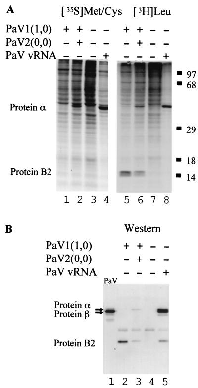
FIG. 3.
Viral proteins synthesized in BSR-T7/5 cells transfected with PaV cDNA clones. (A) In vivo labeling. Cells were mock transfected (lanes 3 and 7) or transfected with 100 ng of vRNA (lanes 4 and 8), or 2.5 μg of PaV1(1,0) (lanes 1 and 5), or 2.5 μg each of PaV1(1,0) + PaV2(0,0) (lanes 2 and 6). After 48 h of incubation at 28°C, cells were preincubated for 30 min with medium lacking either methionine and cysteine (lanes 1 to 4) or leucine (lanes 5 to 8), and then proteins were metabolically labeled for 1 h with [35S]methionine-cysteine (lanes 1 to 4) or [3H]leucine (lanes 5 to 8). Cytoplasmic extracts were harvested, resolved by SDS-PAGE on 12.5% gels, and visualized by fluorography. Proteins α and B2 are indicated on the left, and the migration positions of molecular mass markers are shown on the right. (B) Western blot analysis. Samples 5 to 8 from panel A were also resolved by SDS-PAGE on a 12.5% mini-gel (lanes 2 to 5) along with 250 ng of purified wild-type PaV virus particles (lane 1). Proteins were transferred to a polyvinylidene difluoride membrane, probed with a rabbit antiserum raised against purified PaV particles, and visualized by chemiluminescence.

