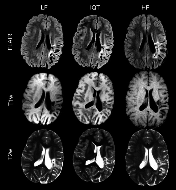
Fig. 3.
LF, IQT-enhanced and HF images from a 15-year-old patient with long-standing encephalomalacic damage in the middle cerebral artery territory on the left from previous perinatal ischemia. The malacic area is better seen on T2w and FLAIR images, while the partial voluming in IQT on T1w images makes the brain cortex look thick. In this case, the 3D visualisation allowed by IQT, even though advantageous compared to the original 2D images, is not completely accurate and may have negative consequences on lesion identification, which stresses the importance of having multiple sequences

