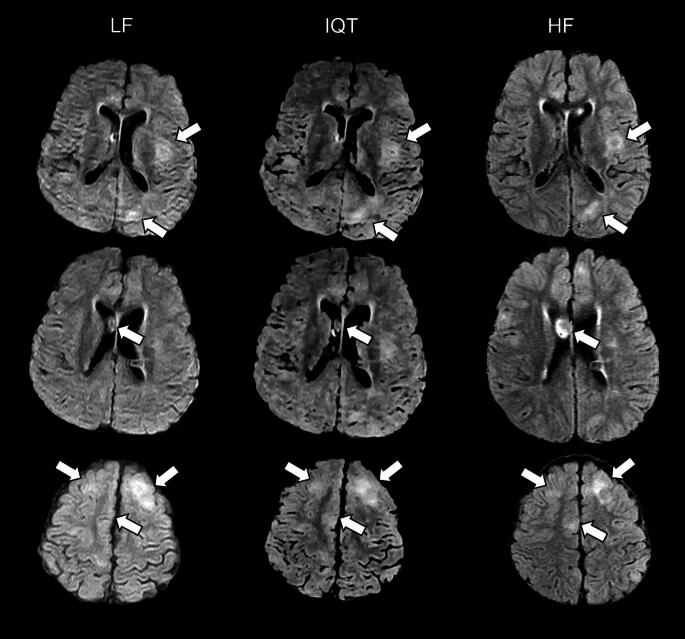
Fig. 4.
LF, IQT-enhanced and HF images from a 10-year-old patient with tuberous sclerosis. Three different levels are shown in the three rows, with tuberous lesions marked by arrows (cortical dysplasias in the upper and lower rows, sub-ependymal nodule in the middle row). Please note that HF images were acquired in a slightly different orientation than LF images, so the HF slices shown here are as close as possible to the LF and IQT-enhanced ones, but not perfectly matched. The location and extension of the tubers are better appreciated on IQT than on the LF scan. This is critical in epilepsy lesion identification for surgical workup

