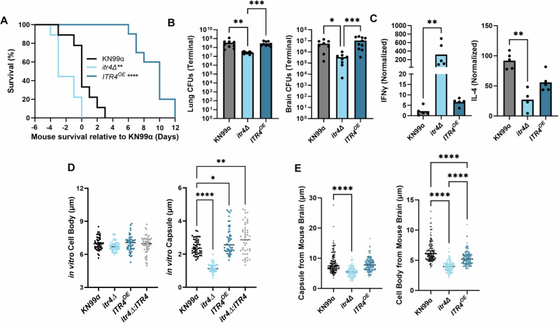
Fig. 6. ITR4OE is attenuated with lower IFNγ than itr4Δ.
A Mice were infected with 5 × 10⁴ cells of itr4Δ, ITR4OE, and KN99α. Significance was determined using a two-sided Gehan-Breslow-Wilcoxon test (n = 10 mice; itr4Δ P = 0.0472; ITR4OE P < 0.0001). B Lungs and brain were collected at terminal endpoints. Significance was determined using a Kruskal–Wallis nonparametric test with Dunn’s multiple comparison correction (n = 10 mice; Lung: from left to right: P = 0.0012, P = 0.001; Brain, from left to right: P = 0.0233; P = 0.006). C Mice were sacrificed on day 17–20 post infection and lungs removed. The lung supernatant was collected, and cytokine levels determined. Fold-change was calculated relative to an uninfected control. Significance was determined using a Kruskal–Wallis nonparametric test (n = 5 mice; IFNγ: itr4Δ P = 0.0016; IL-4: itr4Δ P = 0.0065). D KN99α, itr4Δ, ITR4OE, and itr4Δ:ITR4 were grown for three days in media supplemented with DME, stained with India ink, and imaged (n = 50; Capsule, from left to right: P < 0.0001; 0.0189; 0.0017). E Mice were sacrificed at terminal endpoint and lungs and brains were removed. Cryptococcal cells were collected from each organ, stained with India ink, and imaged. Capsule and cell body size from at least 50 cells per mouse from 5 mice were measured from the brain. Significance was determined using ordinary one-way ANOVA (n = 150; Capsule, from left to right: P < 0.0001; Cell body, from left to right: P < 0.0001; P < 0.0001; P < 0.0001). Source data are provided as a Source Data file. (**P < 0.01; ****P < 0.0001).

