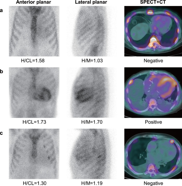
Figure 2 Representative cases of PYP imaging at 3 h after the tracer administration.
A: The anterior and lateral planar images show Grade 1 uptake. No significant myocardial uptake was observed on SPECT. The H/CL ratio at 3 h was 1.58 (recommended cutoff value of ≥1.3), indicating a false-positive result due to the left ventricular cavity uptake of PYP. The H/M ratio at 3 h was 1.03, which was lower than our proposed cutoff value of 1.19.
B: The anterior and lateral planar images show Grade 3 uptake. Significant myocardial PYP uptake was observed on SPECT imaging. Both H/CL and H/M ratios were higher than the cutoff values.
C: The anterior planar image shows Grade 1 uptake; however, the lateral planar image shows Grade 3 uptake. No significant myocardial uptake was observed on SPECT. The H/CL and H/M ratios showed borderline values for the detection of myocardial PYP uptake.