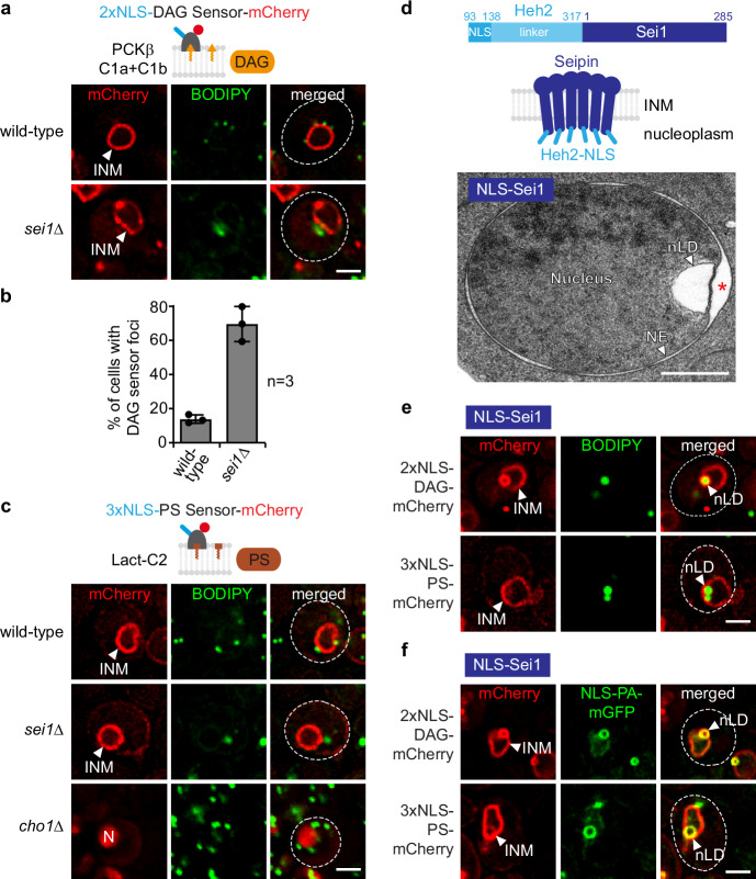
Fig. 3. Seipin differentially alters lipid dynamics at the INM.
a Live imaging of wild-type or sei1∆ cells expressing plasmid-based 2xNLS-DAG-mCherry sensor and stained with BODIPY. INM, inner nuclear membrane. Scale bar, 2 μm. b Quantification of cells with 2xNLS-DAG-mCherry sensor foci in (a). Mean value and standard deviation indicated. n, number of biological replicates. 454 cells for sei1∆ and 628 cells for wild-type analysed. Source data are provided as a Source Data file. c Live imaging of wild-type, sei1∆ or cho1∆ cells expressing plasmid-based 3xNLS-PS-mCherry sensor and stained with BODIPY. INM, inner nuclear membrane; N, nucleus. cho1∆ cells were supplemented with ethanolamine and the sensor was expressed from the GPD promoter in cho1∆ cells. Scale bar, 2 μm. d Cartoon of the engineered NLS-Sei1 construct which contains the nuclear localization sequence (NLS) and the linker of the INM transmembrane protein Heh2 (aa93-317) appended to Sei1. Putative membrane topology of Sei1 is based on cryo-EM models. TEM analysis of a representative example of NLS-Sei1-expressing cells. Plasmid-based mGFP-NLS-SEI1 was expressed from the SEI1 promoter in a sei1∆ strain. N, nucleus; NE, nuclear envelope; nLD, nuclear lipid droplet. Asterisk marks a widened perinuclear space beneath an nLD. Scale bar, 0.5 μm. e Live imaging of sei1∆ cells expressing plasmid-based NLS-SEI1 and lipid sensors tagged with mCherry. Cells are stained with BODIPY. nLD, nuclear lipid droplet; INM, inner nuclear membrane. Scale bar, 2 μm. f Live imaging of sei1∆ cells expressing plasmid-based NLS-SEI1, NLS-PA-mGFP and lipid sensors tagged with mCherry. nLD, nuclear lipid droplet; INM, inner nuclear membrane. Scale bar, 2 μm.

