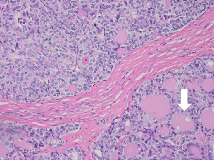
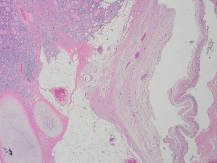
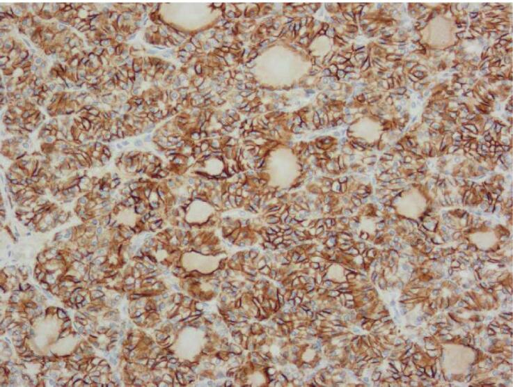
Fig. 1.
Histopathology findings. A: Hemotoxylin and Eosin (H&E) stain X20 magnification: Mature cystic teratoma with proliferative thyroid tissue, consistent with struma ovarii. B: H&E ×20 magnification: Abnormal proliferation of the struma ovarii component with small glands and infiltrative borders. C: H&E X200 magnification: Neoplastic proliferation of thyroid tissue forming follicles with ground-glass nuclei and colloid in the lumen. D: H&E X400 magnification: Neoplastic proliferation of the thyroid tissue forming follicles with ground glass type nuclei with colloid in the lumen. E: TTF-1 Immunohistochemistry X20 magnification: Neoplastic struma ovarii cells show positive nuclear staining. F: Thyroglobulin immunohistochemistry ×200 magnification: Neoplastic struma ovarii cells show positive membrane staining.


