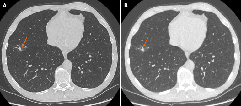
Figure 1.
Representative images demonstrating image quality and ability to identify pulmonary nodules on both standard dose computed tomography chest and ultra-low-dose computed tomography chest with model-based iterative reconstruction imaging protocols. A: Selected axial slice of a standard dose computed tomography (CT) chest presented in lung windows with a solid pulmonary nodule with spiculation and pleural tethering in the lateral segment of the middle lobe (arrow); B: Selected axial slice of an ultra-low dose CT chest in the same patient at the same level presented in lung windows with the same correctly identified pulmonary nodule in the middle lobe (arrow). These images demonstrate the ability of ultra-low-dose CT chest with model-based iterative reconstruction to adequately maintain diagnostic accuracy with regard to solid pulmonary nodules.

