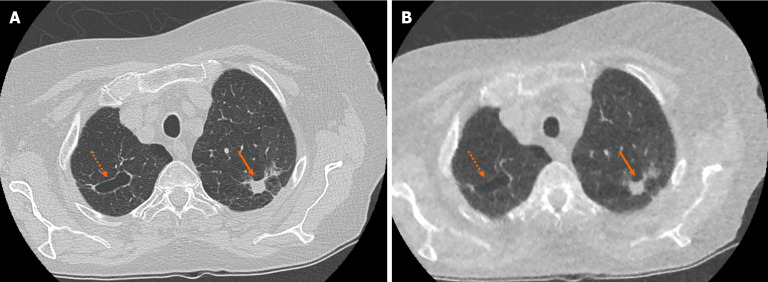
Figure 2.
Example of accurate pulmonary nodule characterisation on ultra-low-dose computed tomography chest. A: Selected axial slice of a standard dose computed tomography (CT) chest presented in lung windows with a spiculated solid pulmonary nodule with pleural tethering in the apico-posterior segment of the left upper lobe (solid arrow) and a parenchymal cyst in the apical segment of the right upper lobe (dashed arrow); B: Selected axial slice in the same patient presented in lung windows at the same level highlights the ability of ultra-low-dose CT chest with model-based iterative reconstruction to correctly characterise pulmonary nodule features such as spiculation, tethering and cavitation.

