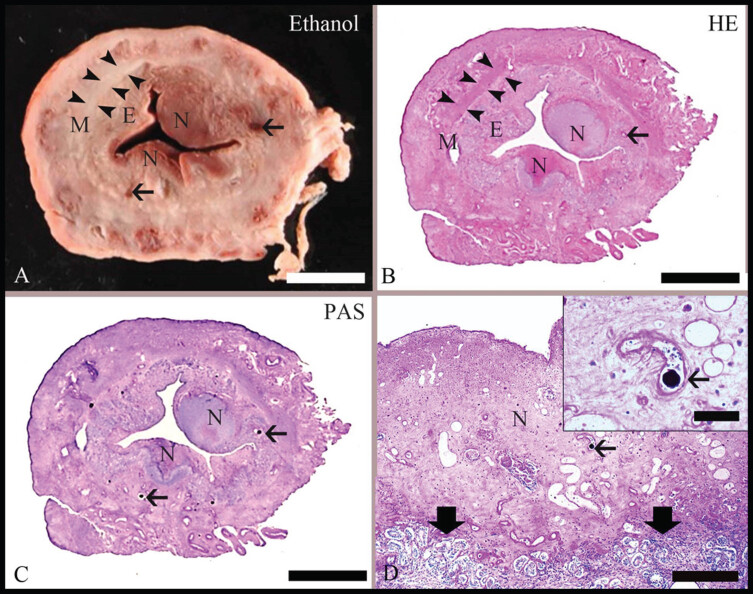
Fig. 1.
Pathomorphological findings in the uterus after embolization with degradable starch microspheres (DSMs). (A): Representative slice from a uterine horn with ethanol fixation (Ethanol-Fix.) used for the scoring of uterine necrosis. On gross examination, the inner endometrium [E] can be distinguished from the surrounding myometrium [M, with segments labeled by arrowheads]; areas of endometrial necrosis [N] are characterized by brownish discoloration. The thin arrows indicate blood vessels. Bar = 0.5 cm. (B): Microscopic hematoxylin–eosin (HE)-stained section corresponding to the formalin-fixed part of the uterine slice illustrated in (A). By microscopic examination, the distinction between E and M as well as the presence of N was confirmed. Bar = 0.5 cm. (C): Microscopic section stained with the periodic acid–Schiff reaction [PAS] and corresponding to the ethanol-fixed part of the slice illustrated in (A). A few vessels contain PAS-positive globular material [arrows], which is consistent with the DSMs. Bar = 0.5 cm. (D): A representative PAS-stained section at a higher magnification. The area of necrosis [N] is characterized by loss of normal tissue histology. It is distinct [thick arrowheads] from the unaffected endometrium. An intralesional vessel with intraluminal PAS-positive globular material is labeled with an arrow. Bar = 200 μm. Inset: Close-up of an intralesional vessel that contains PAS-positive DSM [arrow]. Bar = 50 μm.

