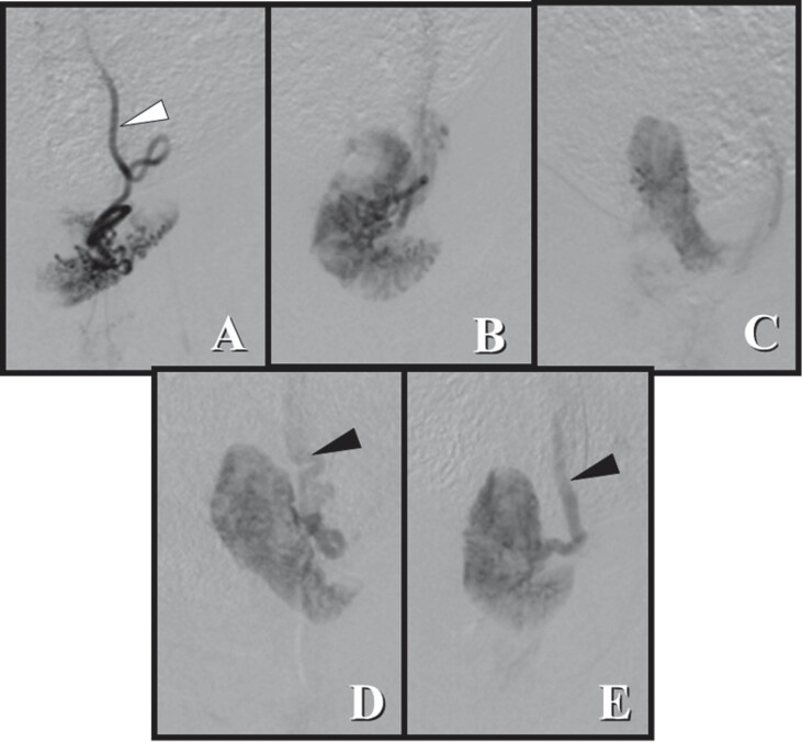
Fig. 4.
Angiographic evaluation of the uterine horn before and after embolization. (A) After 24 hours, there was an almost complete reduction in parenchymal staining. (B) After 48 hours, partial parenchymal staining was observed, but some areas were still not perfused. (C) After 72 hours, parenchymal staining was almost completely restored, and (D) after 7 days, there was complete recovery of homogenous parenchyma compared with the image before embolization. (E). The outline of the uterine parenchyma varied due to uterine horn mobility. White arrow: Uterine artery; black arrow: uterine vein.

