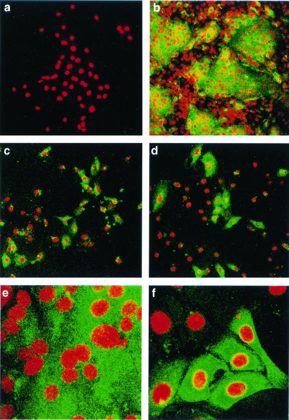
FIG. 4.
Detection of IBV in infected Vero cells by indirect immunofluorescence. Vero cells at 60% confluency were infected with IBV, fixed after 18 h with 50% methanol-acetone, analyzed by indirect immunofluorescence with rabbit anti-IBV polyclonal sera, followed by FITC-labeled goat anti-rabbit antibody, and then stained with propidium iodide to visualize the nuclear DNA. (a) Vero cells that had been infected with Beau-R and were analyzed with preimmune rabbit serum. The remaining panels show Vero cells, analyzed with rabbit anti-IBV serum, infected with Beau-US, exhibiting syncytium formation (b and e); Beau-CK (c); or Beau-R (d and f). Magnifications: a to d, ×16; e and f, ×63.

