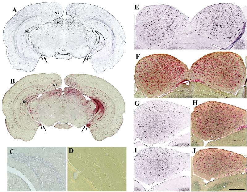
FIG. 3.
Symmetrical and sustained expression from AAV2-HβH following intraventricular injections at birth as shown by in situ hybridization (A, C, E, G, and I) and enzyme histochemistry (B, F, H, and J). Symmetrical pattern of positive cells at the level of the caudal forebrain-rostral midbrain at 1 month p.i. (A and B). The relative positions between the interpeduncular cistern (arrow) and ventral dentate gyrus (arrowhead) are shown. The dentate gyrus of injected brain was negative with a sense riboprobe (C). The dentate gyrus of normal, uninjected C3H mice does not produce detectable levels of enzyme activity (D). (E to J) AAV2 vector expression in the inferior colliculus. Numerous cells were positive for human GUSB at 1 (E and F), 6 (G and H), and 12 (I and J) months p.i. Scale bars: 1.25 mm (A and B), 250 μm (C and D), and 500 μm (E to J). Abbreviations: DG, dentate gyrus; SC, superior colliculus. See Fig. 1 legend for other abbreviations.

