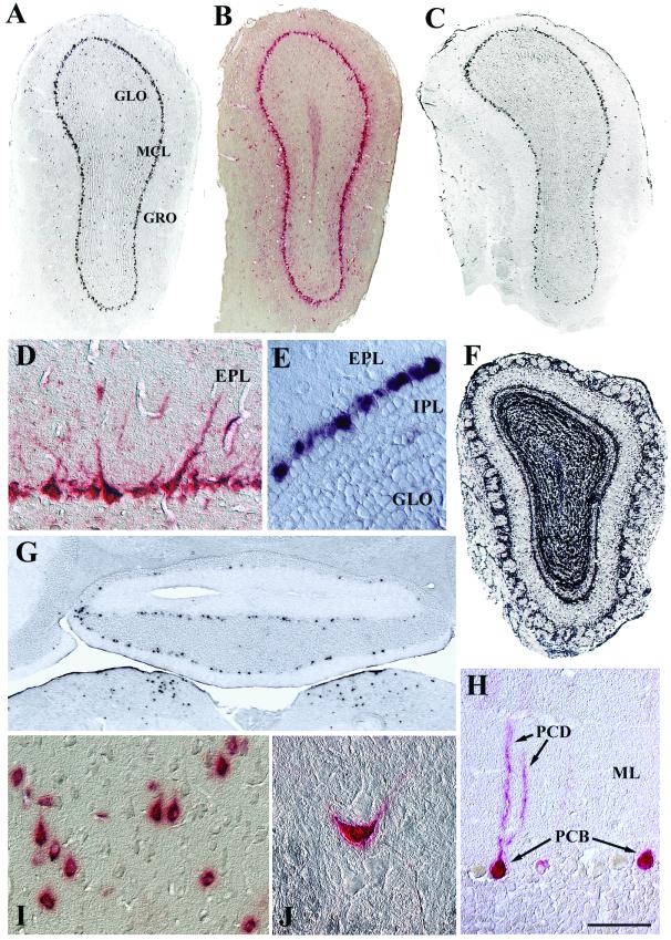
FIG. 5.
Transduction of principal neuron layers as shown by in situ hybridization (A, C, E, F, and G) and enzyme histochemistry (B, D, and H to J). AAV2-HβH expression was detected throughout the MCL at 1 (A and B) and 12 (C, D, and E) months p.i. In addition, a small number of positive cells were scattered in the granule and glomerular layers (A to C). (F) An uninjected transgenic mouse showed that expression of the human GUSB promoter was not restricted to mitral cells. (G) Expression in the medulla oblongata (lower part of panel) and Purkinje cell layer of the cerebellar cortex at 1 month p.i. (H) Purkinje cell morphology was evident with enzyme histochemistry at 12 months p.i. (I) In the motor cortex at 6 months p.i., many enzyme-positive cells possessed pear-shaped somas, characteristic of pyramidal cell morphology. (J) The ventral horn at 12 months p.i. showed an enzyme-labeled neuron in a field of unlabeled neurons. Scale bars: 500 μm (A to C, F, and G) and 50 μm (D, E, and H to J). Abbreviations: EPL, external plexiform layer; GLO, olfactory granule cell layer; GRO, olfactory glomerular cell layer; IPL, inner plexiform layer; ML, molecular layer of the cerebellum; PCB, Purkinje cell bodies; PCD, Purkinje cell dendrites.

