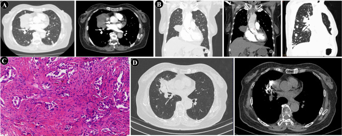
Fig. 1.
Radiological and pathological findings during diagnosis and treatment of pulmonary adenocarcinoma. (A) Chest CT on August 24, 2021: Occupying lesion at the right hilum of the lung, with multiple nodules of varying sizes in both lungs. (B) Percutaneous biopsy guided by CT. (C) Pathological of the percutaneous biopsy (×100). (D) Final follow-up chest CT on August 12, 2023: The lesion at the right hilum of the lung has decreased in size and stabilized, with visible aggregation of radioactive iodine-125 particles within the lesion, while metastatic foci remain indistinct

