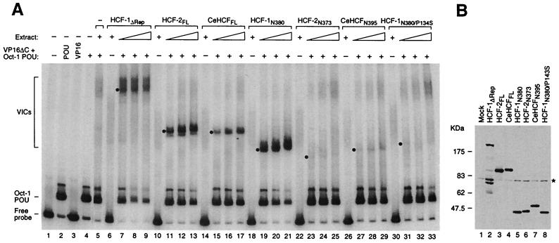
FIG. 2.
All three HCF proteins can stabilize the VP16-induced complex. (A) VP16-induced-complex formation. COS cell extracts containing different HCF proteins were analyzed for VP16-induced-complex formation activity with VP16ΔC, the Oct-1 POU domain, and radiolabeled VP16 response element DNA probe, as described in Materials and Methods. Protein-DNA complexes were resolved in an electrophoretic mobility retardation assay. The positions of the free probe, the Oct-1 POU domain-bound probe, and the VP16-induced complexes (VICs) formed by different HCF proteins are indicated on the left. Lane 1, probe alone; lane 2, probe with Oct-1 POU domain; lane 3, probe with VP16ΔC. Lanes 4, 5, 7 to 9, 11 to 13, 15 to 17, 19 to 21, 23 to 25, 27 to 29, and 31 to 33 contain the Oct-1 POU domain and VP16ΔC. Lane 5 contains in addition unprogrammed COS cell extract, and lanes 6 to 33 contain the COS cell extract with HCF protein as indicated. Each set of three titration lanes contains a twofold titration, and the non-VP16-containing sample contains the most concentrated extract. The position of each VP16-induced complex is indicated with a dot. +, present; −, absent. (B) Immunoblot analysis of the COS cell extracts used in the electrophoretic mobility retardation assay. Extracts containing HA-tagged HCF proteins were resolved by sodium dodecyl sulfate–8% polyacrylamide gel electrophoresis, transferred to a nitrocellulose membrane, and blotted with the 12CA5 anti-HA antibody. The asterisk on the right indicates a nonspecific band. The multiple species smaller than 175 kDa in lane 2 are unrelated to HCF-1 but instead reflect 12CA5-specific cross-reacting cellular proteins that appear only in this sample because it contains the most cell extract after sample normalization.

