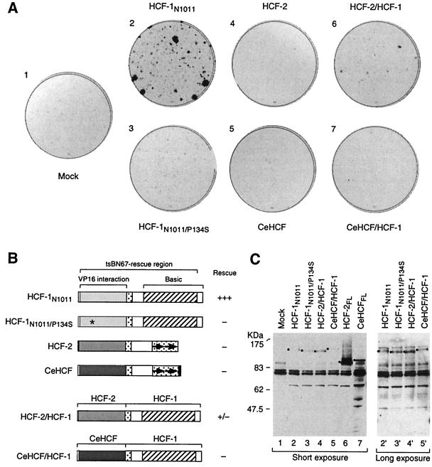
FIG. 4.
The HCF-2KEL repeat region can participate in rescue of the tsBN67 cell proliferation defect. (A) Colony formation assay. tsBN67 cells were transfected with different HCF expression plasmids as indicated. After transfection, the plates were incubated at 40°C for 14 days to permit colony formation. The colonies were visualized by staining them with crystal violet. (B) Schematic diagrams of HCF proteins analyzed and relative levels of rescue of tsBN67 cell proliferation at 40°C. See the legend to Fig. 1A for a description of the diagrams. Asterisk, P134S tsBN67 point mutation. (C) Levels of HCF proteins in transfected tsBN67 cells at nonpermissive temperature. Transfected cells (E plates [see Materials and Methods]) were collected 48 h after transfection and temperature shift to the nonpermissive temperature (40°C) and were used to make protein extracts. The extracts were resolved by sodium dodecyl sulfate–8% polyacrylamide gel electrophoresis, transferred to a nitrocellulose membrane, and probed with the 12CA5 anti-HA epitope tag antibody to monitor the levels of HA-tagged HCF protein synthesis. The position of each relevant band is indicated with a black dot. A long exposure of the immunoblot is shown for lanes 2 to 5 (lanes 2′ to 5′).

