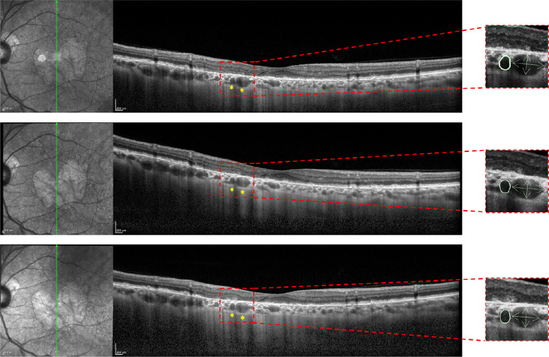
Figure 2.
Example of the grading made to obtain OCT metrics of the choroidal vessels. For each patient, choroidal vessels were identified in the three regions of interest. In this particular case, two choroidal vessels were found within the GA area at baseline and were monitored throughout the follow-up visits. As shown on the right side of the image, measurements were taken for each vessel, including the area, as well as the horizontal and vertical diameters separately, at each visit. Before starting the measurements, the grader determined the vessel's orientation, ensuring that the vertical and horizontal diameters were measured in alignment with the vessel's respective orientation (i.e., the vertical diameter aligned with the vertical orientation of the vessel).

