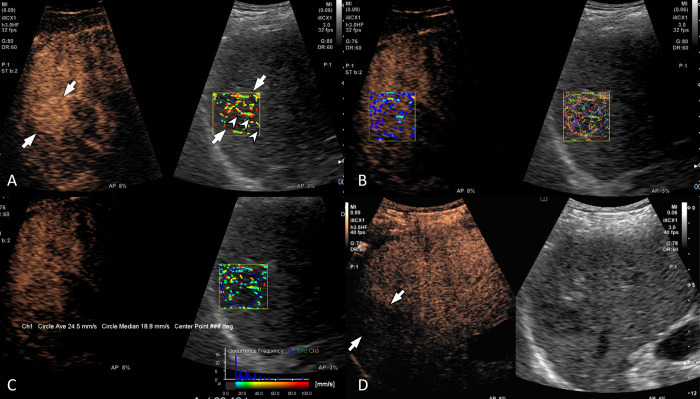
Fig 3. A 63-year-old man with hepatitis C viral infection-related liver cirrhosis and a 3 cm hepatocellular carcinoma in the liver segment 7.
(A) Contrast-enhanced ultrasound (left) and velocity mode of contrast vector imaging (CVI) (right) show a diffuse staining pattern (arrows) during the arterial phase. A penetrating vessel (arrowheads) is well visualized on CVI. (B) Both velocity variance mode (left) and trace mode (right) of CVI show a diffuse staining pattern in the arterial phase. (C) Histogram analysis showed variable velocity and the mean velocity was 24.5 mm/s. (D) The lesion (arrows) showed a late mild washout pattern.

