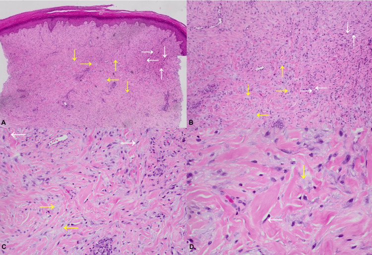
Figure 5. Skin biopsy with hematoxylin and eosin stain.
A. Low power view; B. Low power view; C. Medium power view; D. High power view
Spindle cell proliferations are variably arranged in fascicles composed of relatively bland, elongated cells (white arrows) with areas of interstitial vacuolated material with a faint blue hue resembling mucin (yellow arrows).

