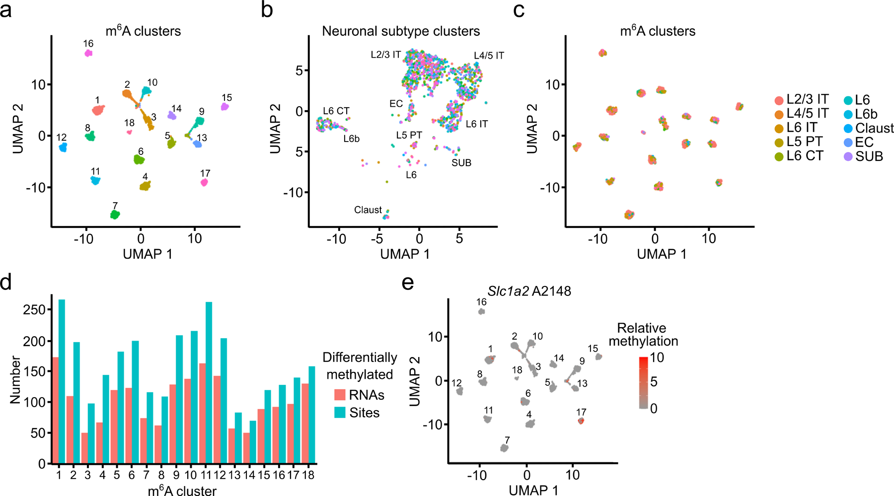
Fig. 5 – Distinguishing cells based on their m6A profiles.

a, UMAP visualization of glutamatergic neurons clustered by m6A methylation (n = 2,246). b, UMAP visualization of glutamatergic neurons clustered by gene expression but colored by m6A cluster (n = 2,246). c, UMAP visualization of glutamatergic neurons clustered by m6A but colored by neuronal subtype (n = 2,246). d, Number of differentially methylated sites and RNAs identified within each m6A cluster. e, UMAP visualization of relative %C2U values of A2148 in the Slc1a2 mRNA within individual cells of m6A clusters (n = 2,246). A2148 is the most significantly differentially methylated site in Slc1a2.
