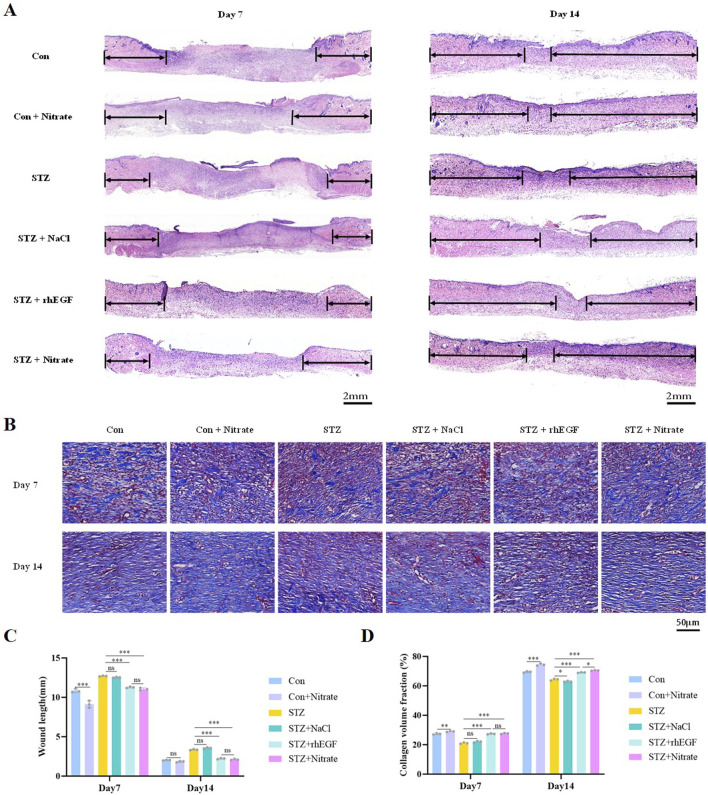
FIGURE 3.
Dietary nitrate promoted re-epithelialization and collagen deposition. (A, C) H&E staining analysis was performed on wound sections at day 7 and 14 post-wounding, with arrows indicating areas of epithelialization. Scale bars = 2 mm. (B, D) Masson’s trichrome staining was conducted at day 7 and 14 post-wounding to assess collagen volume fraction. Scale bars = 50 μm. n = 3, * P < 0.05, ** P < 0.01, *** P < 0.001.

