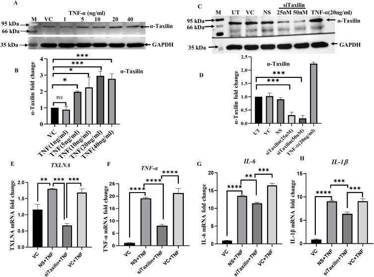
Figure 2.
TNF-α induction upregulates α-Taxilin and inflammation in SW982 synovial cells. (A) The upper panel showing WB images of α-Taxilin after cells were treated with different doses (1, 5, 10, 20, and 40ng/ml) of TNF-α along with its respective GAPDH loading control (lower panel). (B) The graph represents the fold change of α-Taxilin based on the normalized densitometric value of WB upon different doses of TNF-α. (C) The panel showing WB images of α-Taxilin after transfection with different doses (25nM and 50nM) of Si-RNA along with GAPDH loading control (lower panel). (D) The bar graph represents the fold change of α-Taxilin expression based on densitometric values of WB after knockdown using 25nM and 50nM Si-RNA induction and 20ng/ml TNF-α induction along with non-specific Si-RNA control. (E) The α-Taxilin mRNA levels were calculated based on qRT-PCR and represented as a bar graph, α-Taxilin was found to be significantly downregulated when transfected with 25nM/ml siTaxilin. (F) The graph showing the fold change of TNF-α mRNA expression in different treated groups. (G) The graph showing fold change of IL-6 mRNA expression in different treated groups. (H) The graph showing fold change of IL-1β mRNA expression in different treated groups. (* P ≤0.05, ** P ≤0.01, *** P ≤0.001, **** P ≤0.0001).
Abbreviations: WB, Western-blot; ns, Non-significant; NS, non-specific siRNA; TNF-α, Tumor necrosis factor-α; IL-6, Interleukin-6; IL1β, Interleukin-1-β; VC, Vehicle Control; UT, Untreated; kDa, kilo Dalton; GAPDH, Glyceraldehyde 3-phosphate dehydrogenase; nM, Nano molar; ng, Nano gram; mL, milliliter.

