Fig. 4.
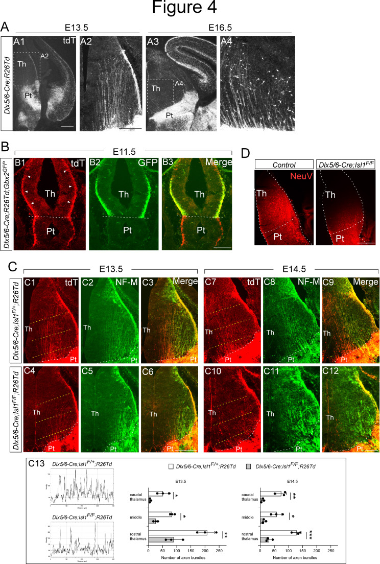
Isl1 is required for the formation of prethalamo-thalamic pioneer axons. (A) Immunohistochemistry for tdTomato on coronal sections of Dlx5/6-Cre; R26-tdTomato embryos at E13.5 and E16.5. Prethalamo-thalamic axons (PTAs) labeled by tdTomato extended into the thalamus, forming ordered and parallel projections. (B) Costaining for tdTomato and GFP on coronal sections of Dlx5/6-Cre; R26-TdTomato; Gbx2GFP embryos at E11.5. PTAs labeled with tdTomato extended into the thalamus, while GFP+ thalamic axons had yet to enter the prethalamus. (C) Costaining for tdTomato and NF-M on coronal sections of control (Dlx5/6-Cre; Isl1F/+:R26-tdTomato) and mutant (Dlx5/6-Cre; Isl1F/F:R26-tdTomato) embryos at E13.5 and E14.5. Loss of Isl1 caused a visible reduction in the number of tdTomato+ axons projecting from the prethalamus to the thalamus in E13.5 and E14.5 mutant embryos. Control NF-M+ thalamic axons formed ordered and parallel projections, but mutant NF-M+ thalamic axons aggregated into thick bundles that ran laterally. (C13) Examples of the measurements taken for the quantification. Yellow dashed lines in C1, C4, C7, C10 indicate the position of the lines used to quantify the number of axon bundles crossing the thalamus. (two-way ANOVA with Sidak’s multiple comparison test; *p < 0.05, **p < 0.01, ***p < 0.001). (D) Implanting the chemical tracer Neurovue into the prethalamus of control (Dlx5/6-Cre; Isl1F/+) at E13.5 visualized PTA projections across the thalamus. Coronal sections from five independent experiments revealed a defective pattern in the outgrowth of PTAs in mutant embryos (Dlx5/6-Cre; Isl1F/F) similar to that shown by tdTomato immunohistochemistry in Dlx5/6-Cre; Isl1F/F; R26-TdTomato embryos. Pt, prethalamus; Th, thalamus. Scale bars: 200 μm

