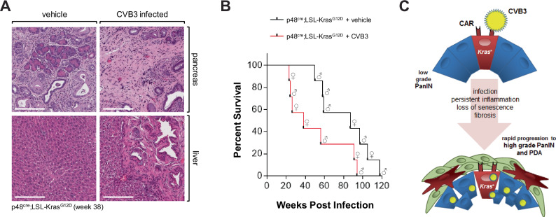
Fig. 6. CVB3 infection of KC mice leads to rapid progression to PDA.
A H&E staining of representative pancreas and liver tissue from (n = 4) control or CVB3 infected p48cre;LSL-KrasG12D mice. Tissue was collected at week 38 after infection. The bar indicates 200 µm. B Kaplan–Meier curve showing significantly reduced survival in n = 7 CVB3-infected p48cre;LSL-KrasG12D mice (red line) as compared to n = 7 PBS control p48cre;LSL-KrasG12D (black line, p = 0.0471; Log-rank (Mantel–Cox) test. The median survival of KC + vehicle is 87 weeks, and the median survival of KC + CVB3 is 38 weeks. Sex of animals is indicated by the symbols. Source data are provided as a Source Data file. C Model of how CAR in oncogenic KRas expressing lesions facilitates CVB3 infection, and how this can contribute to the progression to high-grade PanIN and PDA.

