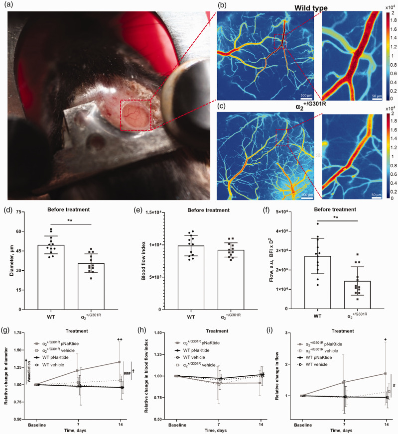
Figure 1.
Increased cerebrovascular tone in mice was prevented by pNaKtide, an inhibitor of the Na,K-ATPase-dependent Src signaling. Representative image of a head-fixed awake mouse with a chronic cranial window (a). Representative laser speckle contrast images from a wild type (WT; b) and a mouse before treatment (c). The diameter of the 3rd order branch of the middle cerebral artery (MCA) was reduced in awake mice prior to treatment compared with WT mice (d; n = 12). The MCA blood flow index (BFI), which is proportional to velocity, did not differ between genotypes (e), with reduced diameter being associated with a lower blood flow (BFI*D2; a.u., arbitrary units) in MCAs from mice before the treatment compared with that of WT (f). Arterial diameter was increased in mice after 14 days pNaKtide treatment (g; n = 6). The pNaKtide treatment did not change arterial diameter in WT mice (n = 6), and vehicle-treated mice of both genotypes (n = 6) also showed no change in diameter over time. The velocity was not statistically different after pNaKtide treatment or vehicle treatment in any of the genotypes (h). The 14 d pNaKtide treatment increased blood flow in mice but did not change blood flow in pNaKtide-treated WT, nor and WT mice receiving vehicle (i). ** indicates P < 0.01 for comparison between genotypes before treatment; +, ++ indicate P < 0.05, and 0.01 for comparison of the effect of 14 d pNaKtide treatment with baseline values in mice; #, ### indicate P < 0.05, 0.001, comparing and WT mice after 14 d pNaKtide treatment; † indicates P < 0.05, for pNaKtide-treated compared with vehicle-treated mice. Data, mean ± SD. Data was compared with two-way ANOVA followed by Bonferroni post-test for multiple comparisons.

