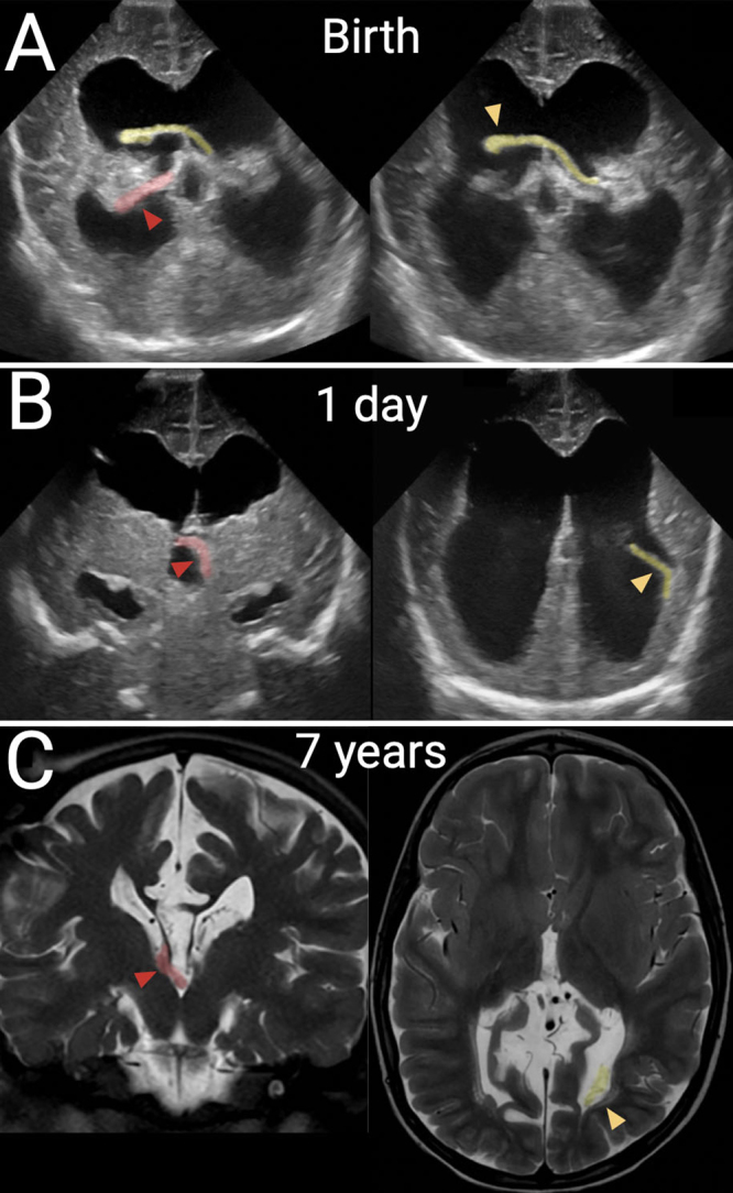
FIG. 4.

Second case of dangling choroid. A full-term patient was born with myelomeningocele that was repaired and treated with a shunt on the 1st day of life. In all panels, the right-sided glomus is shaded in red with a red arrowhead; the left-sided glomus is shaded in yellow with a yellow arrowhead. A: Prior to shunting, coronal transfontanelle ultrasound images show displacement of a mobile left glomus across the perforated septum and a right glomus draped over the posterior thalamus. B: After shunting, coronal ultrasound images show a dangling left glomus in the left ventricle and a newly displaced right glomus in the third ventricle. C: T2-weighted MRI at 7 years of age in the same patient, showing stable locations of both ChPs as compared to the postshunt ultrasound image.
