
Figure 2.
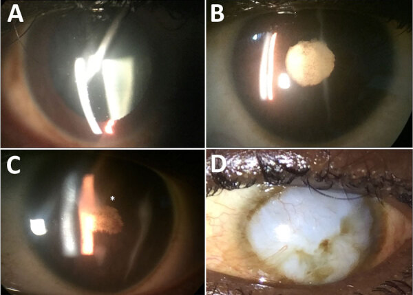
Anterior segment photographs showing the spectrum of ophthalmic sequelae associated with EVD in survivors, Sierra Leone. A) A patient with anterior uveitis has diffuse round keratic precipitates on the corneal endothelium, predominantly within the inferior cornea. B) An EVD survivor with severe, chronic uveitis has posterior synechiae, pigment on the lens capsule, and a dense cataract. C) Another EVD survivor with severe uveitis has dense posterior synechiae overlying a cataract, leading to blindness, and corneal edema involving the superior paracentral cornea (asterisk). D) An external photograph shows a diffuse corneal opacity (leukoma) with superior neovascularization, which was not present before the onset of acute EVD. EVD, Ebola virus disease.