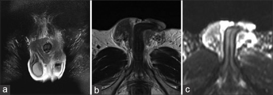
Figure 2.

Magnetic resonance imaging findings. T2-weighted images of the penis revealed heterogeneous lesions with indistinct margins surrounding the penis. The mass was predominantly of low signal intensity (a and b). The apparent diffusion coefficient indicates no diffusion-limited areas within the mass (c)
