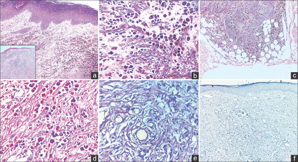
Figure 2.
a: Histopathology image disclosing nodular proliferation of spindled and epithelioid pigmented melanocytes (H & E,40x). Inset: Low-power silhouette of tumor; b: The tumor cells are heavily pigmented and have abundant eosinophilic cytoplasm, vesicular nuclei and prominent nucleoli (H & E,400x); c: The tumor cells are seen extending to the subcutaneous fat (H & E,100x); d: PAS-D stain negative for mucin (400x); e: Melanin bleached H & E image (400x); f: Ki-67 IHC stain displaying low proliferative index (<1%) in the tumor proper (100x)

