
Figure 1.
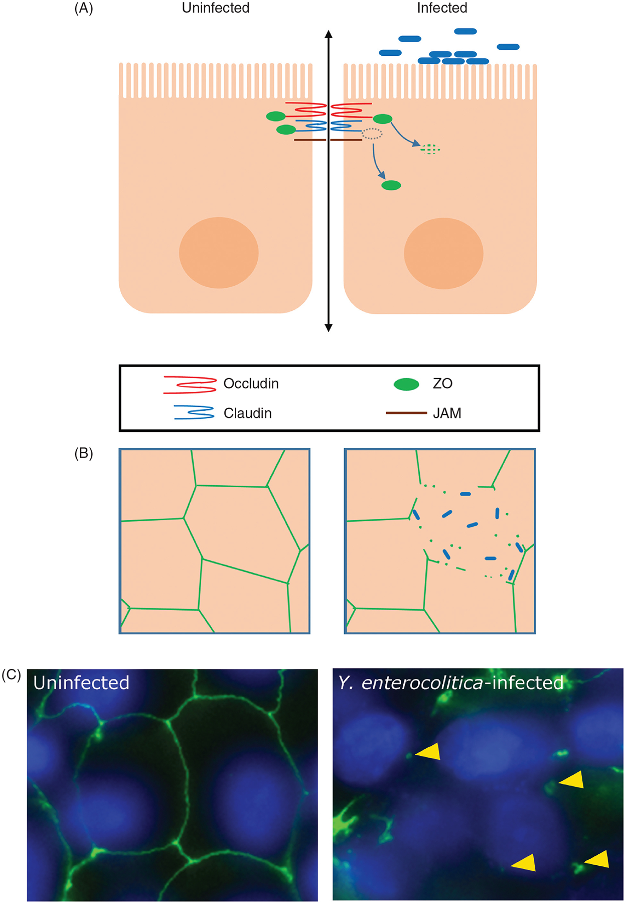
Depletion and relocalization of TJ proteins and TJ-associated proteins disrupts epithelial barrier. (A) Schematic, lateral view: Pathogens may relocalize TJ proteins (occludin, claudins, JAM, and ZO proteins) to intracellular sites and/or trigger their loss. (B) Schematic, en face view: Intact peripheral staining (green) of junctional proteins in uninfected cells. Junctional staining is lost and TJ protein aggregates form in the cytoplasm of infected cells. (C) Example: ZO-1 (green) is preserved at the cell junctions of cultured human intestinal epithelial C2BBe cells. In cells infected with Yersinia enterocolitica, ZO-1 junctional staining is lost, and cytoplasmic ZO-1 aggregates (yellow arrowheads) are observed. DAPI (blue) stains DNA. Image obtained, with permission, from Jennifer Lising Roxas and V.K. Viswanathan.