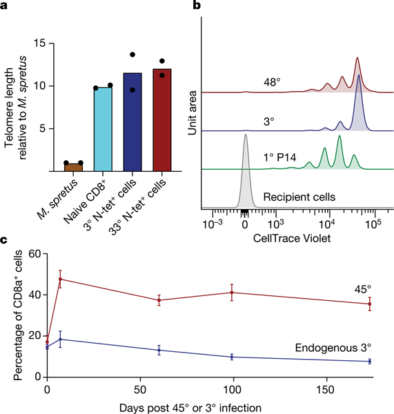
Fig. 2 |. Iteratively boosted T cells maintain telomere length, cell cycle control and durability.
a, Naive, 3° memory or 33° memory CD8+ T cells were sorted, and then tested for telomere length by quantitative PCR and compared to Mus spretus reference DNA. b, 3° and 48° cells were labelled with CellTrace Violet division-tracking dye, and then transferred to recipients without further boosting. Primary lymphocytic choriomeningitis virus (LCMV)-specific P14 memory CD8+ T cells were transferred for comparison. Cumulative cell divisions, indicated by dye dilution, were evaluated in spleen 34 days later. c, 45° and endogenous 3° memory CD8+ T cells were tracked in blood for 173 days following infection. a–c are representative of two experiments with similar results, n = 2 (a), n = 4 (b), n = 5 (c). Error bars show average and s.e.m. For flow cytometry gating strategies, see Supplementary Fig. 2.

