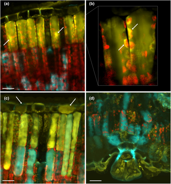
Fig. 2.

Inter‐ and intra‐cellular distribution of flavonoids (FLAV) and hydroxycinnamic acid derivatives (HCAs) in 6‐month‐old Phillyrea latifolia leaves newly developed in full sunlight. Cross sections were stained with Naturstoff reagent (NR, phosphate‐buffered (pH 6.8) saline (1%, w/v, NaCl) solution of 0.1% (w/v) 2‐amino ethyl diphenyl boric acid) and merged fluorescence images (a–d) result from confocal laser scanning microscopy (CLSM) analysis under the following, sequential, excitation (exc)/emission (em) setups. λexc = 365/λem = 415–485 nm for HCA‐derived blue fluorescence; λexc = 488/λem = 565–535 nm for FLAV‐derived yellow fluorescence; λexc = 638/λem = 690–785 nm for chlorophyll‐derived red fluorescence. FLAV accumulate in the vacuoles and the nuclei of adaxial parenchyma (arrows in a), in the outer envelope membranes of the chloroplasts (arrows in b), and in the vacuoles of adaxial epidermal cells (arrows in c). HCAs occur in abaxial mesophyll cells, which have a palisade‐like morpho‐anatomy (as typically occurs in sun‐adapted leaves), together with yellow fluorescent FLAV (d). The multicellular glandular trichome exclusively accumulates FLAV in the vacuole and likely in the cytoplasm, whereas HCAs are merely distributed in the wall of the trichome stalk cell (d). Bars, 20 μm.
