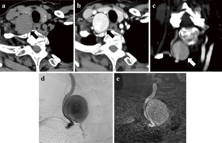
Fig. 2.
Axial images of plain computed tomography demonstrating a mass lesion (arrow) (a). Axial (b) and coronal (c) images of a computed tomographic angiogram demonstrating a contrast-enhanced aneurysmal lesion (arrow). (d) Pre-embolization angiography showing the giant aneurysm originating from the V1 portion of the right VA. (e) Vaso CT. The diameter of the aneurysm is about 30 mm, and its neck is 7.6 mm.

