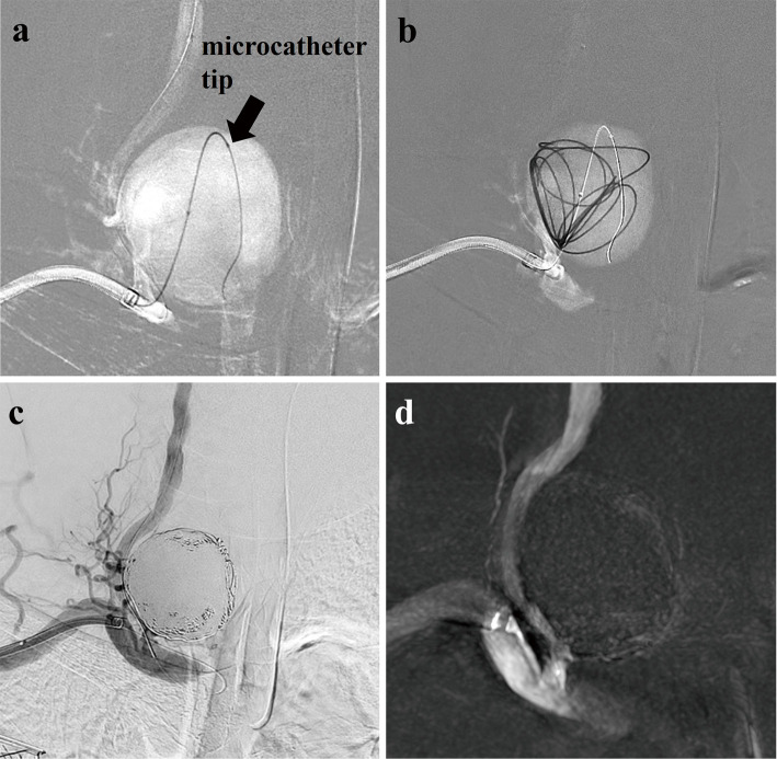
Fig. 3.
Fluoroscopic X-ray images showing that the tip of the microcatheter (arrow) is positioned on the distal side of the aneurysm cavity, (a) and the first coil is inserted into the aneurysm as a frame (b). Postembolization angiography (c) and Vaso CT (d) showing almost complete occlusion of the aneurysm cavity with low-level blood flow in the aneurysm neck.

