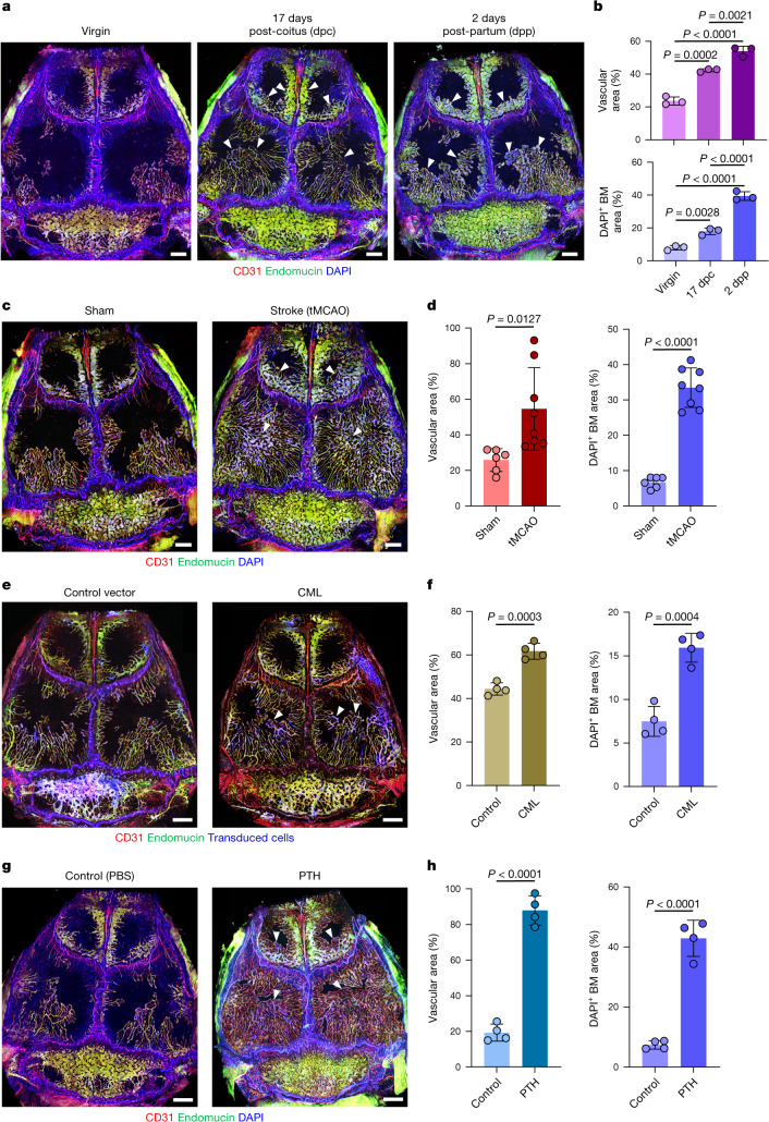
Fig. 2. Pathophysiological regulation of vessel growth and BM expansion in adult skull.
a,b, In vivo immunofluorescence (a) and quantification (b) of skull blood vessels and BM in pregnant (17 dpc) and postpartum (2 dpp) female mice. n = 3 mice per group from 3 independent experiments. c,d, In vivo immunofluorescence (c) and quantification (d) of skull blood vessels and BM in mice 7 days after transient mid-cerebral artery occlusion. n = 6 (sham) and n = 8 (tMCAO) mice per group from 3 independent experiments. e,f, In vivo immunofluorescence (e) and quantification (f) of skull blood vessels and BM in mice with CML. n = 4 mice per group from 3 independent experiments. g,h, In vivo immunofluorescence (g) and quantification (h) of skull blood vessels and BM in mice with 28-day sustained PTH treatment. n = 4 mice per group from 3 independent experiments. Arrowheads indicate areas of substantial expansion. Scale bars, 1 mm. Data are mean ± s.d. P values by Tukey multiple comparison test (one-way ANOVA) and two-tailed unpaired Student’s t-test.

