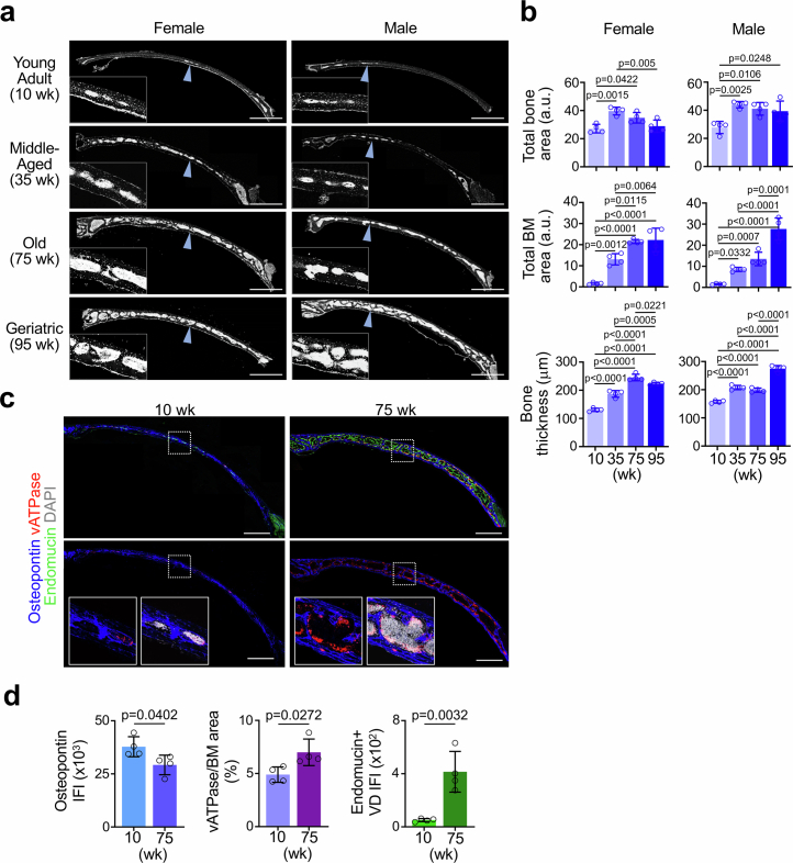
Extended Data Fig. 1. Age-dependent expansion of BM in skull.
a, b. DAPI staining and quantification of mouse skull coronal cryosections showing expansion of BM cellular content during adulthood and aging (n = 4 mice/group from three independent experiments). Blue arrowheads indicate location of magnified inset. c, d. IF staining and quantification of skull coronal cryosections showing increased vATPase+ activated osteoclasts attached to Osteopontin+ bone surfaces in old versus young specimen (n = 4 mice/group from two independent experiments). Scale bars, 1 mm. Vertical bars indicate mean ± SD. P values were calculated using Tukey multiple comparison test (one-way ANOVA) and two-tailed unpaired Student’s t-test.

